
tabulation types
The patterns formed by the alveolae (without or containing thecal plates), the tabulation, have been used to describe and distinguish dinoflagellate taxa for a long time. Dinoflagellates can be athecate (= without thecal plates — "empty" or amorphous material containing amphiesmal vesicles — also named naked or unarmoured), or thecate (= with cellulosic plates in the amphiesmal vesicles, also named armoured) or pelliculate (= having a dino-pellicle as principal strengthening layer of the amphiesma). Six basic types of tabulation have been recognized and are used to classify the taxa: gymnodinioid, suessioid, peridinioid, gonyaulacoid, dinophysoid, and prorocentroid. Detailed descriptions are given in, for example, Fensome et al. (1993) and Hoppenrath and Saldarriaga (2008, Tree of Life webpage for dinoflagellates, http://www.tolweb.org/Dinoflagellates/2445).
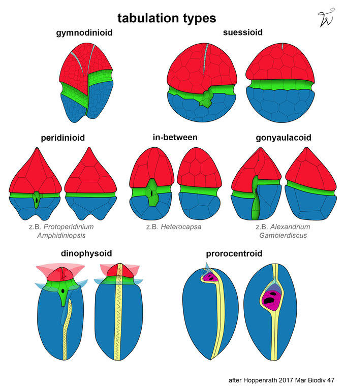
gymnodinioid = Alveolae are numerous and often hexagonal, the cingulum and sulcus being the only clearly distinguishable series. Thecal plates are entirely absent.
suessioid = Alveolae are arranged in 6-11 latitudinal series. The number of plates per series, or even the number of series, varies with species. The cingulum is well marked, and it may contain one or two rows of plates. Thecal plates are delicate.
peridinioid = Alveolae are arranged in five distinct primary latitudinal series. They contain true thecal plates, termed from apex/anterior to antapex/posterior: apicals, precingulars, cingulars, postcingulars, and antapicals. Plates lying between these series are termed intercalaries (anterior or posterior on the epi- or hypotheca respectively), and those lying within the sulcus are sulcals. The midventral epithecal plate often spans both the precingular and apical series. By convention it has been termed the first apical plate. At the apex an apical pore complex (APC) is often present, consisting of an outer (Po) and inner (Pi) pore plate, and a small pre-apical platelet (X, canal plate) is often present. Apical plates are those that contact the APC. Peridinioid tabulations are defined by a more-or-less symmetrical first apical plate and by the presence of two antapical plates.
gonyaulacoid = Alveolae are arranged in five distinct primary latitudinal series. They contain true thecal plates, termed from apex/anterior to antapex/posterior: apicals, precingulars, cingulars, postcingulars, and antapicals. Plates lying between these series are termed intercalaries (anterior or posterior on the epi- or hypotheca respectively), and those lying within the sulcus are sulcals. At the apex an apical pore complex (APC) is often present, consisting of an outer (Po) and inner (Pi) pore plate. Apical plates are those that contact the APC. In gonyaulacoid tabulations the first apical plate is asymmetrical. more ...
dinophysoid = The theca is fundamentally divisible into two halves by a vertical serrated sagittal suture, but a cingulum and sulcus are “superimposed” on it, separating an epitheca and hypotheca. There are small plates on the ventral surface of the epitheca, hypotheca, and in the sulcus around the single large flagellar pore. A simple apical pore is located on the ventral side of the epitheca. The arrangement of the plates varies little within the group. Lists (ridges or extensions of the edge of thecal plates) along the cingulum and sulcus edges may be prominent and developed to an extraordinary degree in some genera, producing very bizarre forms.
prorocentroid = The theca is composed of two large plates, which join along the sagittal suture. A cluster of tiny plates (periflagellar platelets) in a regular arrangement surrounds the two pores. The desmokont flagella arise from one pore. The periflagellar platelets lie principally in an excavation of the right thecal plate. A spine/tooth or protrusions/wings can arise from periflagellar platelets. more ...
SENCKENBERG am Meer
Deutsches Zentrum für Marine Biodiversitätsforschung
PD. Dr. Mona Hoppenrath
Südstrand 44, 26382 Wilhelmshaven
